Helaas is deze beschrijving nog niet in het Nederlands vertaald. U vindt hier dus een Engelse artikelbeschrijving.
Prepared Microscope Slides
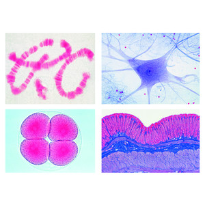
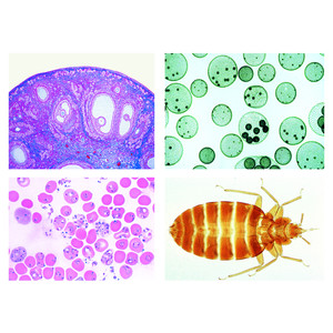
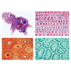
Basic component of the program are the A, B, C and D series comprising of 175 microscope slides. The four series are arranged systematically and constructively compiled, so that each enlarges the subject line of the proceeding one. They contain slides of typical micro-organisms, of cell division and of embryonic developments as well as of tissues and organs of plants, animals and man. Each of the slides has been carefully selected on the basis of its instructional value. LIEDER prepared microscope slides are made in our laboratories under scientific control. They are the product of long experience in all spheres of preparation techniques. Microtome sections are cut by highly skilled staff, cutting technique and thickness of the sections are adjusted to the objects. Out of the large number of staining techniques we select those ensuring a clear and distinct differentiation of the important structures combined with best permanency of the staining. Generally, these are complicated multicolor stainings. LIEDER prepared microscope slides are delivered on best glasses with ground edges of the size 26 x 76 mm (1 x 3"). – Every prepared microscope slide is unique and individually crafted by our well-trained technicians under rigorous scientific control. We therefore wish to point out thatdelivered products may differ from the pictures in this catalog due to natural variation of the basic raw materials and applied preparation and staining methods.
The number of series in hand should correspond approximately to the number of microscopes to allow several students to examine the same prepared microscope slides at the same time. For this reason all slides out of the series can be ordered individually also. So, important microscope slides can be supplied for all students.
School Set B for General Biology
Supplementary Set 50 microscope slides
Zoology and Parasitology
- Paramaecium, nuclei stained
- Euglena, a common flagellate with eyespot
- Sycon, a marine sponge, t.s. of body
- Dicrocoelium lanceolatum, sheep liver fluke, w.m.
- Taenia saginata, tapeworm, proglottids of various ages t.s
- Trichinella spiralis, l.s. of skeletal muscle showing encysted larvae
- Ascaris, roundworm, t.s. of female in region of gonads
- Araneus, spider, leg with comb w.m.
- Araneus, spider, spinneret w.m.
- Apis mellifica, honey bee, mouth parts of worker w.m.
- Apis mellifica, hind leg of worker with pollen basket w.m.
- Periplaneta, cockroach, chewing mouth parts w.m.
- Trachea from insect w.m. - Spiracle from insect w.m.
- Apis mellifica, sting and poison sac w.m.
- Pieris, butterfly, portion of wing with scales w.m.
- Asterias rubens, starfish, arm (ray) t.s. showing tube feet, digestive gland, ampullae
Histology of Man and Mammals
- Fibrous connective tissue of mammal
- Hyaline cartilage of mammal, t.s.
- Adipose tissue, stained for fat
- Smooth (involuntary) muscle l.s. and t.s.
- Medullated nerve fibres, teased preparation of osmic acid fixed material showing Ranvier’s nodes
- Frog blood smear, showing nucleated red corpuscles
- Artery and vein of mammal, t.s
- Liver of pig, t.s. showing well developed connective tissue
- Small intestine of cat, t.s. showing mucous membrane
- Lung of cat, t.s. showing alveoli, bronchial tubes
Botany, Cryptogams
- Oscillatoria, a common blue green filamentous alga
- Spirogyra in scalariform conjugation, formation of zygotes
- Psalliota, mushroom, t.s. of pileus with basidia and spores
- Morchella, morel, t.s. of fruiting body with asci and spores
- Marchantia, liverwort, antheridial branch with antheridia l.s.
- Marchantia, archegonial branch with archegonia l.s.
- Pteridium, braken fern, rhizome with vascular bundles t.s
- Aspidium, t.s. of leaf with sori showing sporangia and spores
Botany, Phanerogams
- Elodea, waterweed, stem apex l.s. showing meristematic tissue and leaf origin
- Dahlia, t.s. of tuber with inuline crystals
- Allium cepa, onion, w.m. of dry scale showing calcium oxalate crystals
- Pyrus, pear, t.s. of fruit showing stone cells
- Zea mays, corn, typical monocot root t.s.
- Tilia, lime, woody dicot root t.s.
- Solanum tuberosum, potato, t.s. of tuber with starch and cork cells
- Aristolochia, birthwort, one year stem t.s.
- Aristolochia, older stem t.s. shows secondary growth
- Cucurbita, pumpkin, l.s. of stem with sieve tubes, annular and reticulate vessels, sclerenchyme fibres
- Root tip and root hairs
- Tulipa, tulip, epidermis of leaf with stomata and guard cells w.m., surface view -
- Iris, typical monocot isobilateral leaf, t.s.
- Sambucus, elderberry, stem showing lenticells and cork cambium, t.s.
- Triticum, wheat, grain (seed) sagittal l.s. with embryo and endosperm


